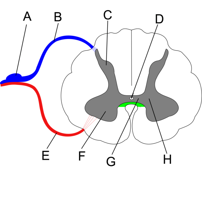
In the realm of neuroscience, the intricate architecture of the spinal cord holds profound significance. Histological analysis of unlabeled spinal cord cross sections provides a unique window into this complex neural landscape, revealing insights into its organization, function, and pathology.
This article delves into the methodologies and applications of this invaluable technique, shedding light on the structural intricacies of the spinal cord.
Unveiling the spinal cord’s intricate architecture through histological analysis of unlabeled cross sections empowers researchers to decipher its intricate organization, function, and pathology. This article provides a comprehensive overview of the methodologies and applications of this invaluable technique, illuminating the structural complexities of the spinal cord.
Histological Analysis of Unlabeled Spinal Cord Cross Sections
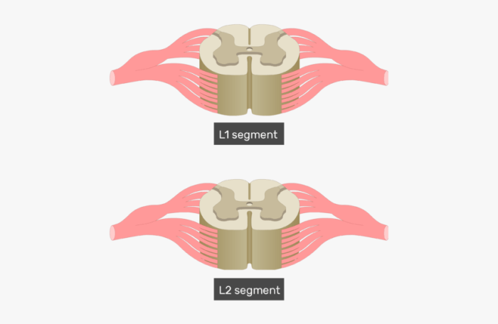
Histological analysis of unlabeled spinal cord cross sections is a valuable technique for examining the morphology and organization of the spinal cord. This analysis allows researchers and clinicians to identify different tissue components, study their distribution, and assess their structural integrity.
Procedure for Preparing and Staining Spinal Cord Cross Sections, Unlabeled spinal cord cross section
Preparing and staining spinal cord cross sections for histological examination involves several steps:
- Tissue fixation:The spinal cord tissue is fixed in a chemical solution (e.g., formalin) to preserve its structure and prevent autolysis.
- Tissue embedding:The fixed tissue is embedded in a support medium (e.g., paraffin or resin) to provide stability during sectioning.
- Sectioning:Thin sections (typically 5-10 micrometers thick) are cut from the embedded tissue using a microtome.
- Staining:The sections are stained with various dyes to enhance the visibility and contrast of different tissue components. Common histological stains include hematoxylin and eosin (H&E), which stains nuclei blue and cytoplasm pink, respectively.
Histological Stains Commonly Used for Spinal Cord Sections
Different histological stains are used to visualize specific components of the spinal cord:
- H&E stain:General purpose stain for visualizing overall tissue architecture.
- Luxol fast blue stain:Stains myelin sheaths blue, highlighting myelinated nerve fibers.
- Cresyl violet stain:Stains Nissl bodies (rough endoplasmic reticulum) in neurons, allowing visualization of neuronal distribution.
- Immunohistochemistry:Uses antibodies to visualize specific proteins or molecules within the tissue.
Immunohistochemical Techniques for Unlabeled Spinal Cord Cross Sections
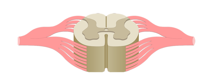
Immunohistochemistry (IHC) is a powerful technique used to visualize and localize specific proteins within tissue sections. It involves the use of antibodies that specifically bind to target proteins, allowing for their detection and analysis. IHC is particularly useful for studying the distribution and expression patterns of proteins in unlabeled spinal cord cross sections, providing insights into cellular and molecular processes within the spinal cord.
Antibody Selection and Optimization
The success of IHC depends on the selection of appropriate antibodies. Antibodies should be specific for the target protein, with minimal cross-reactivity with other proteins. Optimization of antibody concentration and incubation time is crucial to achieve optimal staining intensity and specificity.
This can be done through pilot experiments or by consulting antibody datasheets.
Detailed Protocol for Immunohistochemical Staining
- Tissue Preparation:Prepare spinal cord cross sections using standard histological techniques (e.g., formalin fixation, paraffin embedding).
- Antigen Retrieval:Heat or enzyme treatment may be required to expose hidden epitopes for antibody binding.
- Blocking:Nonspecific binding sites are blocked using a protein-containing solution (e.g., bovine serum albumin).
- Primary Antibody Incubation:Apply the primary antibody diluted in blocking solution to the tissue sections and incubate overnight at 4°C.
- Secondary Antibody Incubation:Incubate with a secondary antibody conjugated to a reporter enzyme (e.g., horseradish peroxidase) to amplify the signal.
- Visualization:Use a chromogenic substrate to visualize the antibody-antigen reaction (e.g., diaminobenzidine).
Examples of Specific Antibodies
- NeuN:Neuronal marker used to identify neurons in the spinal cord.
- GFAP:Glial fibrillary acidic protein, a marker for astrocytes.
- Iba1:Ionized calcium-binding adaptor molecule 1, a marker for microglia.
- Synaptophysin:Presynaptic vesicle protein, used to study synaptic connectivity.
Morphological Assessment of Unlabeled Spinal Cord Cross Sections
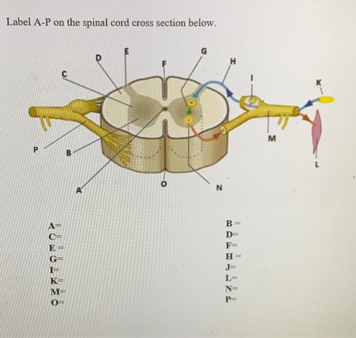
Morphological assessment of unlabeled spinal cord cross sections involves the evaluation of structural characteristics and architectural organization of the spinal cord tissue. This assessment plays a crucial role in understanding the normal organization of the spinal cord and detecting pathological changes associated with neurodegenerative diseases.
Quantitative Morphological Parameters
Various quantitative morphological parameters can be measured from unlabeled spinal cord cross sections, providing insights into cellular and axonal integrity:
- Neuronal Density:The number of neurons per unit area, reflecting neuronal loss or gain.
- Axonal Diameter:The average diameter of axons, indicating axonal damage or regeneration.
- Myelin Thickness:The thickness of the myelin sheath surrounding axons, assessing myelin damage or demyelination.
Significance of Morphological Changes
Morphological changes in the spinal cord can provide valuable information about the underlying pathological processes:
- Neuronal Loss:A decrease in neuronal density may indicate neurodegeneration or cell death.
- Axonal Damage:Reduced axonal diameter or increased axonal swelling can suggest axonal injury or Wallerian degeneration.
- Myelin Damage:Thinning or loss of myelin can indicate demyelination, affecting neuronal function and conduction.
By quantifying these morphological parameters, researchers can gain insights into the extent and severity of spinal cord damage, providing a basis for understanding the underlying mechanisms and potential therapeutic interventions.
Image Analysis and Data Interpretation
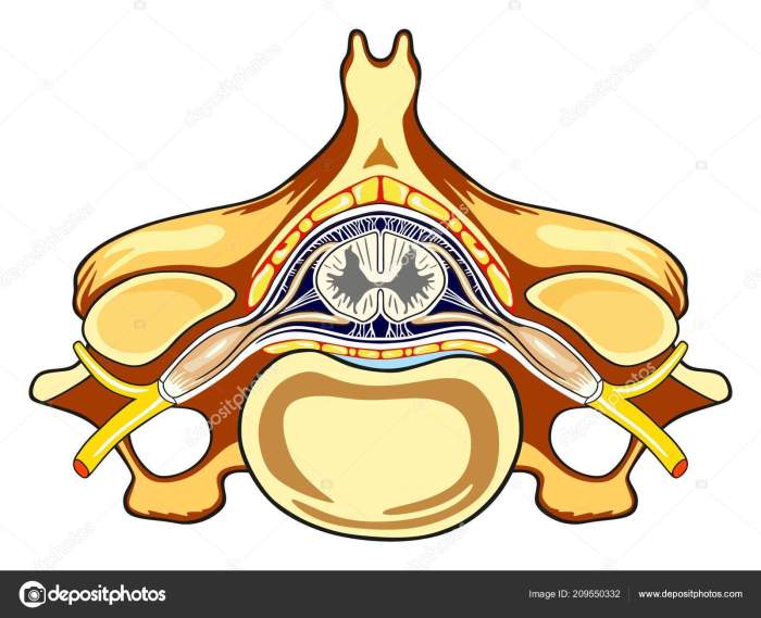
Image analysis is a critical step in the study of unlabeled spinal cord cross sections. It allows researchers to quantify structural and functional parameters of the spinal cord, providing valuable insights into its organization and function.
Several software programs are available for image analysis of spinal cord cross sections, including ImageJ, Fiji, and Imaris. These programs provide a range of tools for image segmentation, feature extraction, and data quantification.
Image Segmentation
Image segmentation is the process of dividing an image into distinct regions or objects. In the context of spinal cord cross sections, segmentation can be used to identify different anatomical structures, such as the gray matter, white matter, and spinal cord meninges.
Feature Extraction
Once the image has been segmented, features can be extracted from each region. These features can include morphometric parameters, such as area, perimeter, and shape, as well as intensity measurements, such as mean pixel intensity and texture.
Data Quantification
The extracted features can then be used to quantify various aspects of spinal cord structure and function. For example, the area of the gray matter can be used to assess the size of the motor and sensory nuclei, while the intensity of the white matter can be used to assess the density of myelinated axons.
Image analysis is a powerful tool that can be used to generate quantitative data on spinal cord structure and function. This data can be used to study the effects of injury, disease, and other factors on the spinal cord.
Applications in Biomedical Research
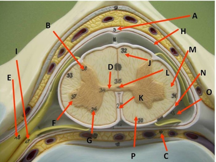
Analyzing unlabeled spinal cord cross sections plays a pivotal role in biomedical research, providing valuable insights into the structure, function, and pathology of the spinal cord.
This technique has been extensively used in studies investigating spinal cord injury, neurodegenerative diseases, and developmental disorders.
Spinal Cord Injury
- Unlabeled cross sections allow researchers to examine the extent of tissue damage, characterize cell loss, and evaluate axonal injury.
- This information is crucial for understanding the mechanisms of spinal cord injury and developing effective treatment strategies.
Neurodegenerative Diseases
- Analysis of unlabeled cross sections helps identify pathological changes associated with neurodegenerative diseases such as amyotrophic lateral sclerosis (ALS) and multiple sclerosis (MS).
- Researchers can study the distribution and progression of lesions, providing insights into disease pathogenesis and potential therapeutic targets.
Developmental Disorders
- Unlabeled cross sections are used to assess spinal cord development during embryogenesis and early postnatal stages.
- This technique enables researchers to identify structural abnormalities, neural tube defects, and other developmental disorders affecting the spinal cord.
The analysis of unlabeled spinal cord cross sections continues to advance our understanding of spinal cord function and pathology, providing a valuable tool for biomedical research and the development of novel therapeutic interventions.
Answers to Common Questions
What are the advantages of using unlabeled spinal cord cross sections?
Unlabeled cross sections provide a comprehensive view of the spinal cord’s overall architecture, allowing for the assessment of various tissue components and their spatial relationships.
How can immunohistochemistry enhance the analysis of unlabeled cross sections?
Immunohistochemistry enables the localization and identification of specific proteins within the spinal cord, providing insights into cellular distribution and function.
What is the significance of morphological assessment in analyzing unlabeled cross sections?
Morphological assessment quantifies structural parameters such as neuronal density, axonal diameter, and myelin thickness, providing valuable insights into the health and integrity of the spinal cord.
