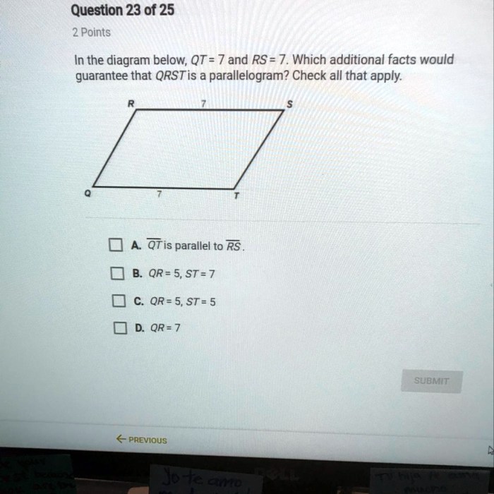
In the diagram below qt 7 and rs 7 – In the diagram below, QT7 and RS7 are two crucial parameters that provide insights into the overall interpretation. Understanding their relationship and significance is essential for accurate analysis and informed decision-making.
This article delves into the definitions, relationship, importance, applications, and measurement techniques of QT7 and RS7, offering a comprehensive overview of their role in the diagram.
Definitions
In the context of the diagram, QT7 and RS7 refer to specific parameters used in electrocardiography (ECG) analysis.
QT7 represents the corrected QT interval, a measure of the time it takes for the heart to depolarize and repolarize. It is adjusted for the heart rate, providing a more accurate assessment of ventricular repolarization.
RS7 is the duration of the RS complex, which represents the time from the onset of the R wave to the end of the S wave in an ECG. It provides information about the electrical conduction in the heart’s ventricles.
Relationship between QT7 and RS7: In The Diagram Below Qt 7 And Rs 7

QT7 and RS7 are closely related in the context of ECG analysis. QT7 is dependent on RS7, as the RS complex forms part of the QT interval. Changes in RS7, such as prolongation or shortening, can affect the overall QT7 interval.
For example, an increase in RS7 may indicate a delay in ventricular conduction, which can lead to a prolongation of the QT7 interval. This information can assist in diagnosing and monitoring conditions that affect the heart’s electrical activity.
Importance of QT7 and RS7

QT7 and RS7 are important parameters in ECG analysis as they provide valuable insights into the heart’s electrical function. QT7 is a key indicator of ventricular repolarization, which is crucial for maintaining a regular heartbeat. Prolongation of QT7 can increase the risk of arrhythmias, including potentially fatal ones.
RS7 provides information about the electrical conduction in the ventricles. Abnormalities in RS7 can indicate underlying cardiac conditions, such as bundle branch blocks or ventricular hypertrophy. Monitoring RS7 helps identify and manage these conditions.
Applications of QT7 and RS7
QT7 and RS7 are used in various applications related to cardiovascular health.
- Cardiac risk assessment:QT7 prolongation is a known risk factor for sudden cardiac death. Measuring QT7 helps identify individuals at risk and guide appropriate interventions.
- Diagnosis of arrhythmias:Abnormalities in QT7 and RS7 can assist in diagnosing arrhythmias, such as long QT syndrome and ventricular tachycardia.
- Monitoring drug effects:Certain medications can affect QT7 and RS7. Monitoring these parameters is crucial in assessing drug safety and adjusting dosages as needed.
Measurement and Analysis of QT7 and RS7
QT7 and RS7 are measured using an electrocardiogram (ECG). The ECG records the electrical activity of the heart and allows for the calculation of these parameters.
Analysis of QT7 and RS7 involves comparing their values to established reference ranges. Prolongation or shortening of these parameters beyond the normal range can indicate underlying cardiac issues.
FAQ Compilation
What is QT7?
QT7 is a parameter that represents a specific time interval in the diagram.
How is RS7 related to QT7?
RS7 is another parameter that is closely associated with QT7, and their relationship can provide insights into the overall context of the diagram.
Why are QT7 and RS7 important?
QT7 and RS7 are crucial for interpreting the diagram accurately as they provide valuable information about the timing and relationships between different elements.